If you are asking what is a cardiac window, the answer depends on the medical context. In many heart-care articles, people use “cardiac window” to mean a pericardial window, which is a surgical opening made in the sac around the heart to drain extra fluid. In heart imaging, a “cardiac window” can also mean an ultrasound window, or the place where a sonographer positions the probe to see the heart clearly.
The most common meaning online is pericardial window. Johns Hopkins describes a pericardial window as a procedure where a small part of the sac around the heart is surgically removed so extra fluid can drain.
Quick Answer: What Is a Cardiac Window?
A cardiac window usually means one of two things:
1. Pericardial window: a surgical drainage procedure for extra fluid around the heart.
2. Echocardiogram window: an ultrasound view used to see the heart during imaging.
If a doctor says someone needs a “window” because there is fluid around the heart, they are usually talking about a pericardial window procedure. If an ultrasound technician says they need a better “window,” they may mean a better imaging angle for the heart.
What Is a Pericardial Window?
A pericardial window is a procedure done on the pericardium, the thin sac surrounding the heart. Normally, the pericardium has a small amount of fluid between its layers to reduce friction as the heart beats. When too much fluid builds up, it is called a pericardial effusion. MedlinePlus describes pericardial effusion as fluid buildup in the sac around the heart, while cardiac tamponade is a serious problem where fluid buildup affects heart function.
During a pericardial window, a surgeon creates an opening so fluid can drain away instead of collecting tightly around the heart. Cleveland Clinic explains that this “window” allows draining fluid to spill into the larger pleural cavity so it does not keep filling the pericardial space.
Why Would Someone Need a Cardiac Window?
A cardiac window may be needed when fluid around the heart is causing symptoms, affecting heart function, or coming back after earlier drainage.
Possible reasons include:
Pericardial effusion
Cardiac tamponade risk
Cancer-related fluid around the heart
Infection
Inflammation of the pericardium
Heart surgery complications
Chest injury
Kidney failure with uremia
Autoimmune disease
Fluid that returns after pericardiocentesis
Johns Hopkins lists several possible causes that may lead to a pericardial window, including infection, cancer, inflammation after a heart attack, injury, immune system disease, medicine reactions, radiation, and kidney failure with uremia.
What Is Pericardial Effusion?
A pericardial effusion means there is too much fluid in the sac around the heart. A small amount of fluid is normal, but too much can create pressure. If pressure becomes severe enough, the heart may not fill and pump properly.
Cleveland Clinic lists symptoms of pericardial effusion that can include shortness of breath, chest pressure or pain, fast heartbeat, palpitations, lightheadedness, fainting, fatigue, and confusion in more serious cases.
Some people have no symptoms at first, especially if the fluid builds slowly. Others may feel chest discomfort, breathing difficulty, dizziness, or weakness.
Cardiac Tamponade: Why Fluid Around the Heart Can Be Dangerous
The main emergency concern is cardiac tamponade. This happens when fluid around the heart builds enough pressure to stop the heart from filling normally. That can reduce blood flow to the body.
StatPearls describes cardiac tamponade as a condition that can present with shortness of breath, chest discomfort, low blood pressure, raised neck veins, muffled heart sounds, and pulsus paradoxus, and says management requires urgent stabilization and prompt drainage through pericardiocentesis or surgery.
A pericardial or cardiac window may be used when doctors need a more lasting way to drain fluid or prevent it from collecting again.
How Is a Cardiac Window Done?
A cardiac window is performed by a trained surgical team. The exact method depends on the patient’s condition, the amount of fluid, the cause, and whether the situation is urgent.
Johns Hopkins explains that a pericardial window may be done under general anesthesia. One approach uses an incision under the bottom of the breastbone, another uses an incision between the ribs, and another may use several small side-chest incisions with video-assisted thoracoscopy, often called VATS.
The goal is to drain the extra fluid and, when needed, send fluid or tissue for testing. That testing can help identify infection, cancer, inflammation, or another cause.
Pericardial Window vs Pericardiocentesis
A pericardial window is not the only way to drain fluid around the heart. Another option is pericardiocentesis, where a needle and thin tube are used to remove fluid from the pericardial sac.
Cleveland Clinic describes pericardiocentesis as a procedure where a needle is inserted into the chest until it reaches the pericardium, allowing doctors to drain fluid directly or place a drain for slower removal.
The difference is simple:
| Procedure | What It Means | Common Use |
| Pericardiocentesis | Needle and catheter drain fluid | Often used for urgent or diagnostic drainage |
| Pericardial window | Surgical opening in the pericardium | Often used for recurrent, difficult, or persistent fluid |
| VATS pericardial window | Minimally invasive surgical window | May be used when a surgical approach is preferred |
Johns Hopkins notes that pericardiocentesis is another way to remove fluid, but if the fluid returns after being drained or the situation makes needle drainage difficult, a provider may be more likely to use a pericardial window.
Is a Cardiac Window Major Surgery?
A pericardial window is a surgical procedure, so it is more involved than simply taking medicine. However, not every case is done through a large incision. Some are done through smaller incisions using VATS, depending on the situation.
The seriousness depends on:
How sick the patient is
Whether tamponade is present
Whether the fluid is caused by cancer, infection, surgery, or trauma
The surgical approach used
The person’s age and overall heart/lung health
Whether the procedure is urgent or planned
For some people, it is done as a planned procedure. For others, it may be part of urgent treatment.
What Symptoms May Lead Doctors to Consider It?
Doctors may investigate fluid around the heart if someone has:
Shortness of breath
Chest pressure or pain
Dizziness or fainting
Fast heartbeat or palpitations
Weakness or fatigue
Trouble breathing when lying flat
Swelling in the legs or abdomen
Low blood pressure in serious cases
Mayo Clinic notes that pericardial effusion symptoms may include shortness of breath, discomfort while lying down, chest pain behind the breastbone or left chest, chest pressure, lightheadedness, and swelling in the legs or belly.
These symptoms can overlap with other heart and lung problems, so testing is needed to confirm the cause.
How Doctors Diagnose Fluid Around the Heart
A doctor may use several tests depending on the situation. The most common heart imaging test is an echocardiogram, also called a heart ultrasound.
StatPearls says pericardial effusion is diagnosed using clinical evaluation and imaging, with echocardiography being the primary method for confirming fluid around the heart.
Other possible tests may include:
ECG/EKG
Chest X-ray
Echocardiogram
CT scan
Cardiac MRI
Blood tests
Fluid testing after drainage
The test plan depends on symptoms, urgency, and suspected cause.
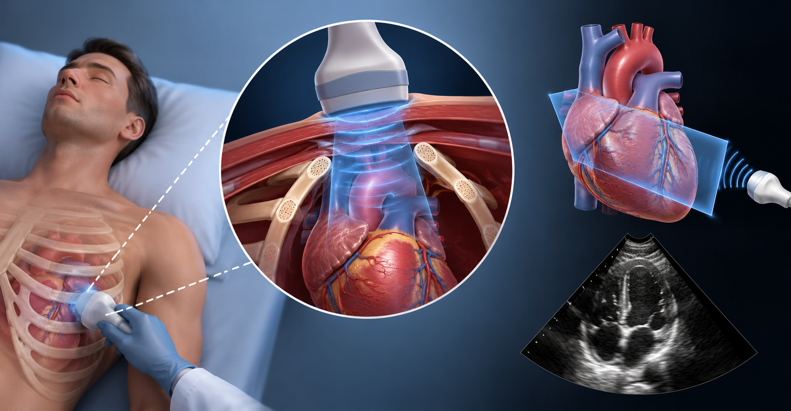
Cardiac Window in Heart Imaging
In imaging, a cardiac window can mean an acoustic window used during an echocardiogram. This is not surgery. It simply means a place where ultrasound waves can pass through the chest well enough to show the heart.
Common echocardiogram windows include:
Parasternal window
Apical window
Subcostal window
Suprasternal window
A medical review of transthoracic echocardiography describes the four standard windows as parasternal, apical, subcostal, and suprasternal.
So if someone says “poor cardiac window” during an echo, they usually mean the images are difficult to obtain because of body shape, lung interference, chest anatomy, surgical dressings, or positioning — not that surgery is needed.
Pericardial Window vs Echo Window
This is where the wording can confuse people.
| Term | Meaning |
| Pericardial window | Surgical opening to drain fluid around the heart |
| Cardiac ultrasound window | Imaging view used during an echocardiogram |
| Poor cardiac window | Ultrasound images are hard to see clearly |
| Subxiphoid window | Can refer to an echo view or a surgical approach, depending on context |
If your report says “limited acoustic window,” that is usually about image quality. If your doctor says “you may need a pericardial window,” that is about draining fluid.
Risks and Possible Complications
Like any procedure, a pericardial window has risks. The exact risks depend on the patient and the surgical approach.
Possible risks may include:
Bleeding
Infection
Pain after surgery
Reaction to anesthesia
Irregular heartbeat
Injury to nearby structures
Fluid returning
Need for another procedure
A surgical team weighs these risks against the danger of leaving fluid around the heart untreated, especially if the effusion is large, recurrent, infected, cancer-related, or affecting heart function.
Recovery After a Pericardial Window
Recovery varies. Some people are monitored in the hospital for a short time, while others need longer care because of the underlying illness. A drain may remain temporarily to help remove fluid. Doctors may also send samples to the lab.
During recovery, the team may watch:
Heart rhythm
Blood pressure
Breathing
Drain output
Pain control
Signs of infection
Whether fluid is returning
The follow-up plan depends on why the fluid developed in the first place.
When to Seek Urgent Medical Help
Fluid around the heart can become dangerous if it affects circulation. Get urgent medical help if someone has:
Severe chest pain
Shortness of breath at rest
Fainting
Blue or grey lips
Confusion
Very fast heartbeat
Low blood pressure symptoms
Sudden weakness or collapse
These symptoms can happen with several serious conditions, including heart attack, pulmonary embolism, severe infection, and cardiac tamponade. They need prompt medical evaluation.
Simple Plain-English Meaning
If you are asking what is a cardiac window, the most common answer is:
A cardiac window usually refers to a pericardial window, a procedure that creates a small opening in the sac around the heart so extra fluid can drain.
In imaging, it can also mean:
An ultrasound window used to view the heart during an echocardiogram.The phrase sounds complicated, but the context tells you which meaning applies. If the topic is fluid around the heart, pericardial effusion, or tamponade, it usually means a drainage procedure. If the topic is echo images, heart ultrasound, or poor window, it usually means an imaging view.